이번 1월, 과학자들은 분자를 처음으로 직접 보았다. 그것도 매우 중요한 분자를.
과학자들은 지난 4백여년동안 너무 작아서 육안으로는 볼수 없는 작은 물체들을 보아왔다.
이런 기술은 처음 렌즈를 사용하면서 시작되었다. 렌즈는 빛을 굴절시키고 이런 방식으로 빛을 반사하는 물체의 상(像)을 모으고(집중) 확대시킬수 있다. 이런 방식을 사용하는 기구가 현미경이었다.
시간이 흐를수록 현미경은 개선되어 마침내 물체를 1천배나 확대시킬수 있게 되었다. 그러나 이 정도에서 과학자들은 물리적 장벽에 부딪쳤다. 빛은 파동들로 구성된다. 파동들은 미세하지만 현미경으로 보는 물체들 역시 미세하다. 물체들이 너무 작다면 그것을 보는데 쓰여지는 빛의 파동보다도 작다는 얘기이다. 이 경우 파동은 물체를 건너뛰게 되어 물체는 볼 수 없게 된다.
이 난관을 피하기위해 자외선같은 보다 짧은 파동을 이용할 수 있다. 그래서 얼마동안 학자들은 '자외선 현미경'을 사용했다. 그러나 이것은 약간의 개선효과만을 가져왔다. 보다 짧은 파동은 적절하게 촛점을 모을수가 없었다.
그러나 1923년 프랑스의 과학자 '루이 드 브로글리'가 원자의 구성 입자들도 파동의 형태로 존재할것이라고 지적했다. 1925년 미국의 과학자 '클린톤 J. 데이비슨'은 전자에 의해 생긴 그러한 파동을 찾아낼수 있었다. 전자파는 보통의 광파보다 훨씬 짧았다. 전자파는 실제 X선파의 크기와 비슷했다. 그러나 X선파는 촛점을 모으기가 대단히 어려웠으나 전자와 그 파동은 자기장으로 쉽게 모을수 있었다.
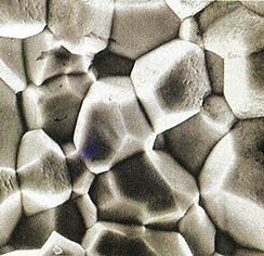
1932년, 이런 방식으로 전자파를 모으고 물체의 상을 확대하는 기구가 독일의 과학자 '에른스트 루스카'에 의해 만들어졌다. 이것이 '전자현미경'이었다. 전자현미경은 처음 조잡했으나 해가 지날수록 다듬어지고 개선되어 마침내 물체를 30만배나 확대시킬수 있었다.
초기에 이런 기구에서 전자는 확대된 상을 얻기위해 물체를 뚫고 들어가야만 했다. 따라서 과학자들은 얇게 썬 물체를 갖고 일을 해야만 했다. 그러나 곧 얇고 섬세한 전자빔을 얻어내고 그 빔을 물체의 표면에 스치게 하는 방법이 발견되었다. 전자빔은 스치는곳을 감지했다. 다시 말하면 표면을 주사(走査)하여 확대된 상을 얻은 것이다.
이것이 '전자 주사 현미경'이었다.
이제 보다 새로운 방식이 등장했는데 이것은 학자들이 '터널링 효과'라고 부르는 방식으로 전자를 끌어내는 것이다. 이리하여 우리는 '전자 주사 터널링 현미경'을 갖게 되었고 이것은 확대기능의 신기록을 세우게 되었다.
이제 이 현미경은 1백만배의 확대기능을 가지며 지난 1월 DNA분자를 최초로 관찰하는데 쓰여졌다. 이때 캘리포니아 '리버모아'연구소의 '마이켈 B. 살메론'과 기타 여러사람이 참여했다.
DNA는 생명의 설계도를 갖고 있기 때문에 중요한 것이다. 모든 살아가는 세포는 자신을 계속 복제하는 DNA 분자 조합을 갖고 있다. 새로 만들어진 분자는 딸세포에 전달된다. 이런 조합은 정자와 난자세포에도 있어 분자가 부모에서 자녀한테로 옮겨질수 있다. 모든 살아있는 종(種)은 자신의 유전자조합을 갖고있으며 같은 종이라도 개체마다 유전자조합에 있어 미세한 차이가 있다.
DNA의 중요성은 1944년에 처음 공인되었고 과학자들은 어떻게 이들 분자들이 자신과 똑같은 분자들을 재생산해 낼 수 있는지를 알아내기 위해 애써왔다.
1953년에 영국의 과학자 '프란시스 H.C.크릭'과 공동연구자인 미국의 '제임스 D.왓슨'은 그것을 밝혀내었다. X선은 분자를 통과할때 옆으로 튀는 경향이 있다. X선의 사진은 X선이 튈때의 점들을 보여준다. 그러한 'X선 회절 양상'으로부터 분자의 모양을 연역해 내는것이 가능하다. 이것은 긴시간과 조심스런 작업을 요했다. 그러나 마침내 DNA분자는 이중나선(침대의 스프링모양)으로 꼬여진 원자들의 두 개의 복잡한 사슬로 구성되었음이 밝혀졌다. 각개의 사슬은 서로 꼭맞게 엉켜있다.
DNA가 자신과 같은 분자를 만들때 두개의 사슬은 풀어지고 각개의 사슬은 세포액에서 약간의 원자군(群)을 취합, 정확히 자신과 똑같은 새로운 사슬을 만든다. 각개의 사슬은 자신의 동반자를 만들때 표준으로 기여한다. 결국 각개의 DNA는 정확히 똑같은 두개의 DNA분자를 만드는 셈이다.
이 연구작업은 '왓슨'과 '크릭'으로 하여금 1962년도 노벨상을 타게했고 치밀한 과학적 연역의 승리로 간주되었다. 두 사람은 DNA분자의 이중나선구조와 그 활동상을 비록 너무 작아 볼수는 없었지만 정확하고 자세하게 기술해 놓은 것이다.
그러나 '왓슨'과 '크릭'의 연구발표가 있은지 36년이 지난 오늘, DNA분자의 그림은 '전자 주사 터널링 현미경'으로 찍혀 나오게 되었으며 치밀한 추리작업은 필요없게 되었다. 눈으로 볼수 있는 이중나선이 있는 것이다. 분자는 추리(상상)한대로 꼬여있다. 그림을 통해 마디사이의 길이로 알아낼수 있다. 그 길이는 약 5백50만분의 1인치이다.
'살메론'과 그의 그룹은 사슬의 더욱 자세한 모양을 보기위해 방법을 개선시킬 계획을 세우고 있다. 그들은 다른 분자들의 상(像)도 얻기위한 노력을 기울일 것이다.
This January, scientists got their first direct look at a molecule, and a very important molecule at that.
Scientists have been looking at objects that were too small to see with the naked eye for nearly 400 years. The trick at first was to use lenses that forced light to bend and in this way focus and enlarge the image of object that reflects the light. The device used to do so was the microscope.
As time went on, microscopes were improved until finally they could magnify objects 1,000 times. At that point, scientists ran up against a physical barrier. Light consists of waves. These waves are tiny, but the objects under the microscope were tiny, too. If the objects were tiny enough, they were smaller than the light waves being used to view them. The light waves then tended to skip over them so the objects could not be seen.
To get around this, you might use shorter light waves such as ultraviolet. For a while, therefore, scientists used "ultra-microscopes," but these represented only a small improvement. Still shorter waves could not be focused properly.
But then, in 1923, a French scientist, Louis de Broglie, pointed out that subatomic particles ought to exist in wave form, too. In 1925, an American scientist, Clinton J. Davisson, was able to detect such waves produced by electrons. These electron-waves were much shorter than ordinary light-waves. They were, in fact, about the size of x-ray waves. But where x-ray waves would be extremely difficult to focus, electrons and their waves could easily be focused by magnetic fields.
In 1932, the first device used to focus electron-waves and enlarge the images of objects in that way was constructed by a German scientist, Ernst Ruska. This was an "electron microscope." It was crude at first, but over the years it was refined and imroved, until it could enlarge objects 300,000 times.
At first, in such instruments, electrons had to pass through an object to produce an enlarged image. Scientists had to work with very thin slices of material. But then, ways were found to produce a very thin, sharp beam of electrons and play that beam over the surface of an object. The beam "felt" its way, so to speak, across the surface, scanning it and producing an enlarged image. This was a "scanning electron microscope."
Now, a still newer version produces the electrons by means of what scientists call a "tunneling effect" and we have a "scanning tunneling electron microscope" that has reached new heights of magnification. It can now magnify 1,000,000 times and was used in January to take a first-ever look at a molecule of DNA by Miquel B. Salmeron and others at the Lawrence Livermore Laboratory in California.
DNA is important because it carries the blueprint of life. Every living cell contains a set of DNA molecules that are constantly replicating themselves, passing the newly created molecules on to daughter cells. Such sets also exist in sperm and egg cells, so that they can be passed on from parents to children. Every living species has its own set and there are also tiny differences in the sets of different individuals of the same species.
The importance of DNA was first recognized in 1944, and scientists labored to find out how these molecules could produce other molecules exactly like themselves.
In 1953, the British scientists Francis H.C. Crick and his American co-worker, James D. Watson, Worked it out. X-rays, in passing through molecules, tend to bounce to the side. Photographs of such X-rays produce dots where the X-rays bounce, and from such "X-ray diffraction patterns" it is possible to deduce the shape of the molecule. It took a long time and careful work, but it finally turned out that the DNA molecule consisted of two complex strands of atoms twined in a double helix (the shape of bedsprings). Each strand had a complicated shape, and the two strands fit each other exactly.
When DNA forms another molecule like itself, the two strands unwind and each one picks up small groups of atoms from the cell fluid and puts them together into a new strand that fits exactly upon the original one. Each strand serves as a model to form a new partner for itself. In the end, then, each DNA molecule produces two DNA molecules exactly alike.
This work brought Watson and Crick the Nobel Prize in 1962, and their work was considered a triumph of subtle scientific deduction. They described the double helix of the DNA molecule and its workings in precise detail, even though it was much too small to actually be seen.
But now, 36 years after the work of Watson and Crick, pictures of a DNA molecule have been taken with a scanning tunneling electron microscope―and no subtle reasoning is required. There is the double helix, visible to the eye. The molecule coils as it is supposed to. From the image, it is possible to work out the distance between successive coils. It is about 5,500,000th of an inch.
Salmeron and his group are planning to refine the method further to see if they can see even finer details of the strands. They will try to obtain images of other molecules, too.
(c) 1989, Los Angeles Times Syndicate
1989년 06월 과학동아 정보
글
함인선 소장글
동아일보사 편집부
🎓️ 진로 추천
- 생명과학·생명공학
- 화학·화학공학
- 물리학

1989년 06월 과학동아 다른추천기사


이 기사를 읽은 분이 본
다른 인기기사는?
다른 인기기사는?

- 과학사에 등장한 4대 악마

- Part 3. 진단부터 치료까지 프랙탈 의학

- Part 3. 로봇의 시대, 엄마로 산다는 것
- [물리]운동량과 운동에너지의 차이점

- Part 5. 온 몸이 보안 코드, 생체인증 시대